Skull Inferior View Worksheet
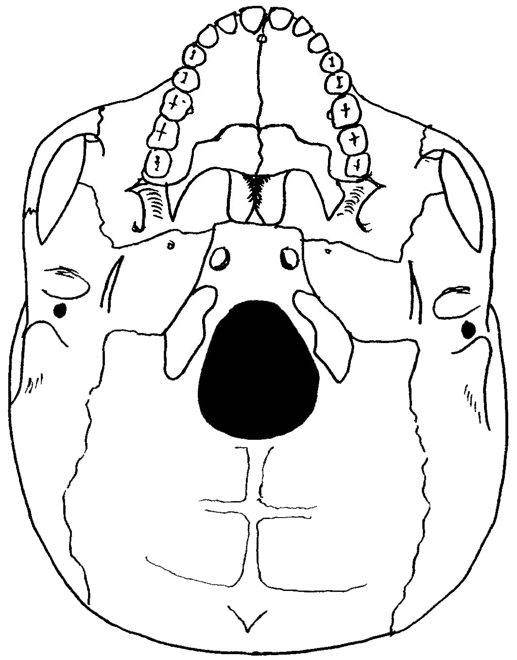
If you're looking for a comprehensive and informative worksheet that focuses on skull anatomy, then the Skull Inferior View Worksheet is exactly what you need. Designed specifically for students and enthusiasts interested in the study of human anatomy, this worksheet provides a detailed exploration of the skull's inferior view, highlighting key structures and their functions.
Table of Images 👆
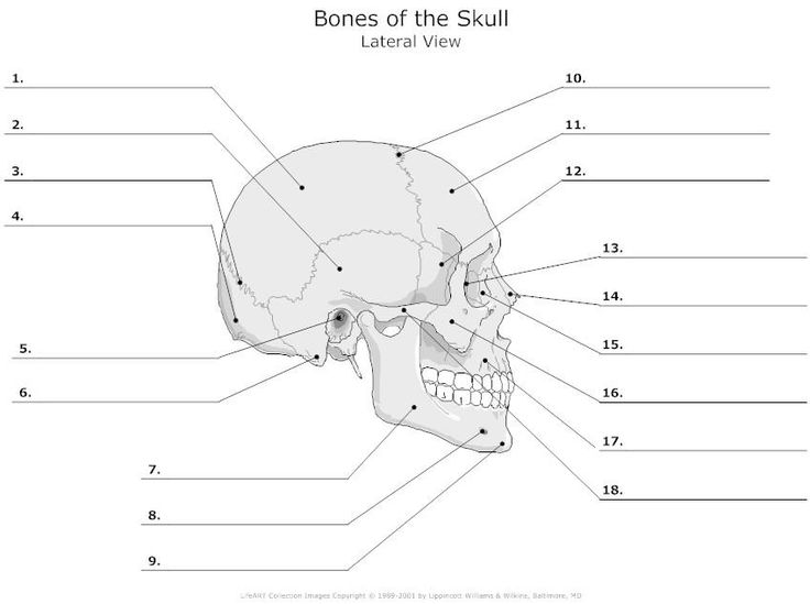
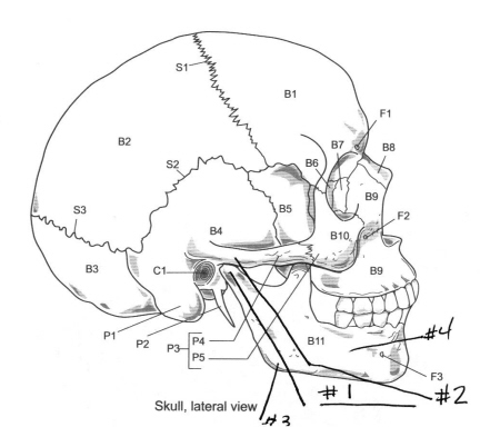
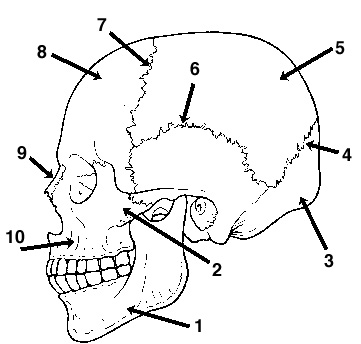
- Skull Bones Anatomy Blank Diagram
- Blank Human Skull Diagrams Inferior View
- Skull Axial Skeleton Labeling Worksheet
- Anatomy and Physiology Bone Worksheets
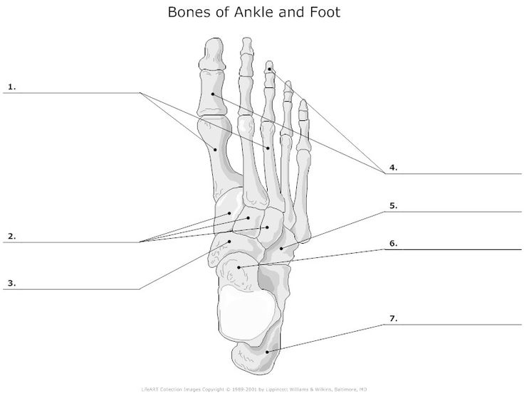
- Foot Bone Diagram Unlabeled
- Axial Skeleton Labeling Worksheets
- Skull Bones and Sutures Quiz
- Blank Skeletal System Diagram Quiz
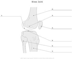
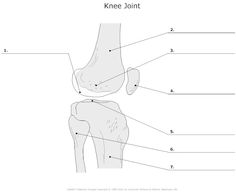
- Unlabeled Knee Joint
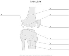
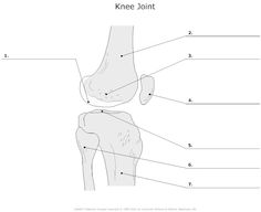
- Unlabeled Knee Joint
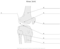
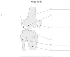
- Unlabeled Knee Joint
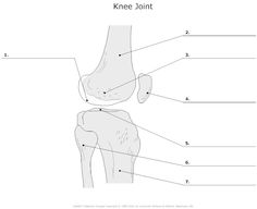
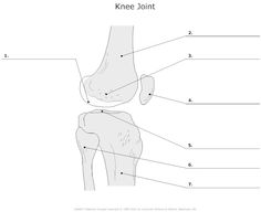
- Unlabeled Knee Joint
- Unlabeled Knee Joint
- Unlabeled Knee Joint
- Unlabeled Knee Joint
- Unlabeled Knee Joint
- Unlabeled Knee Joint
- Unlabeled Knee Joint
- Unlabeled Knee Joint
More Other Worksheets
Kindergarten Worksheet My RoomSpanish Verb Worksheets
Healthy Eating Plate Printable Worksheet
Cooking Vocabulary Worksheet
My Shadow Worksheet
Large Printable Blank Pyramid Worksheet
Relationship Circles Worksheet
DNA Code Worksheet
Meiosis Worksheet Answer Key
Rosa Parks Worksheet Grade 1
What is visible in the inferior view of the skull?
In the inferior view of the skull, you would be able to see structures such as the hard palate, the foramen magnum, the mandible, and the openings of the nasal and oral cavities. Additionally, you may also see parts of the cranial base, including the sphenoid bone and ethmoid bone.
Where is the foramen magnum located?
The foramen magnum is located at the base of the skull, in the occipital bone. It is the largest opening in the skull and serves as a passageway for the spinal cord to connect with the brainstem.
What structures are visible in the occipital bone?
The occipital bone in the skull is known for several key structures, including the external occipital protuberance, the superior and inferior nuchal lines, the occipital condyles that articulate with the first cervical vertebra (atlas) forming the atlanto-occipital joint, the foramen magnum which transmits the spinal cord, and the external and internal occipital crests. These structures play a crucial role in providing support, protection, and articulation within the skull and neck region.
Which bone forms the posterior part of the hard palate?
The bone that forms the posterior part of the hard palate is the palatine bone.
What is the name of the depression that houses the pituitary gland?
The fossa that houses the pituitary gland is called the sella turcica.
Which bone forms the lateral walls of the nasal cavity and part of the orbits?
The ethmoid bone forms the lateral walls of the nasal cavity and part of the orbits.
What bone articulates with the temporal bones to form the temporomandibular joint?
The mandible bone articulates with the temporal bones to form the temporomandibular joint.
What are the three cranial fossae visible in the inferior view?
The three cranial fossae visible in the inferior view are the anterior cranial fossa, middle cranial fossa, and posterior cranial fossa. These fossae house the different regions of the brain and support structures, providing protection and space for various functions within the skull.
Where can the external auditory meatus be found in the inferior view?
The external auditory meatus, also known as the ear canal, can be found in the inferior view on the lateral side of the temporal bone, at the junction of the squamous and mastoid portions of the bone.
What is the name of the structure that separates the nasal and oral cavities?
The structure that separates the nasal and oral cavities is called the palate.
Have something to share?
Who is Worksheeto?
At Worksheeto, we are committed to delivering an extensive and varied portfolio of superior quality worksheets, designed to address the educational demands of students, educators, and parents.











Comments