Structure of the Heart Worksheet
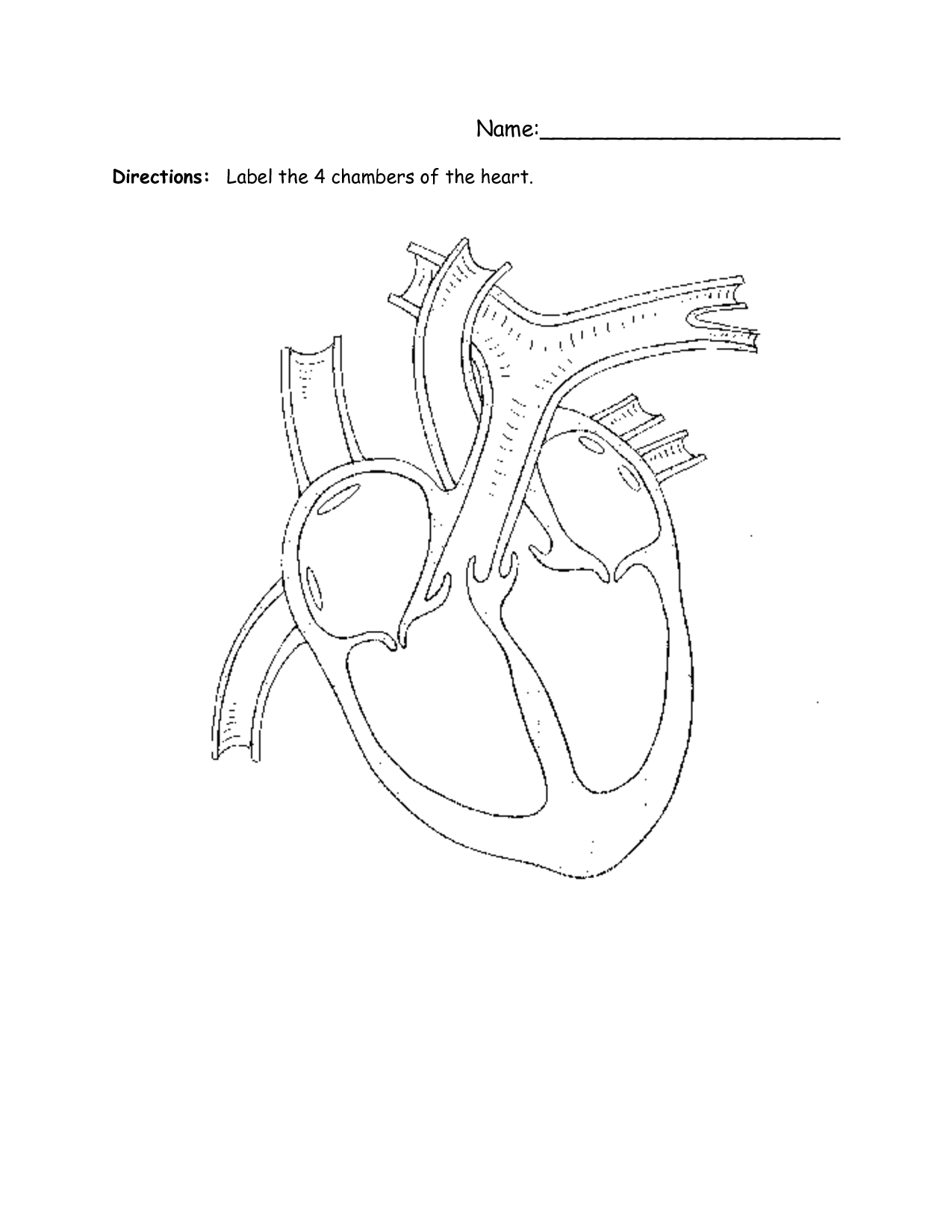
The structure of the heart is a complex and vital topic to understand, especially for students studying anatomy or biology. A worksheet that focuses on this subject can be a great tool for reinforcing the knowledge and identifying the different entity of the heart. By providing clear diagrams and labeling exercises, students can enhance their understanding of the heart's structure in a systematic and organized manner.
Table of Images 👆
- Blank Heart Diagram
- 4 Chambers of the Heart Worksheet Label
- Labeled Animal Cell Worksheet
- Human Brain Diagram Outline
- Basic Atomic Structure Worksheet
- Human Brain Worksheet
- Muscle Movement Worksheet Answers
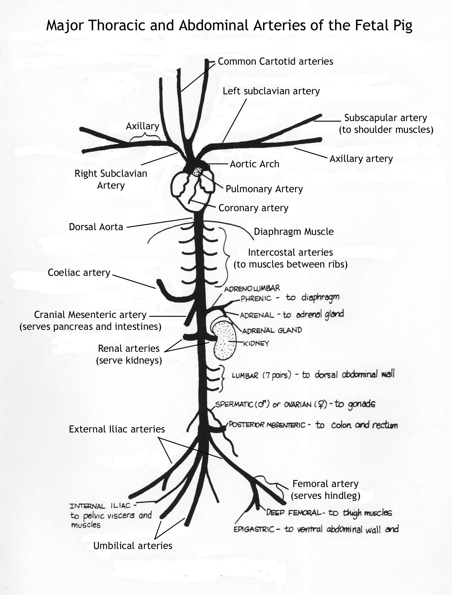
- Fetal Pig Circulatory System Diagram
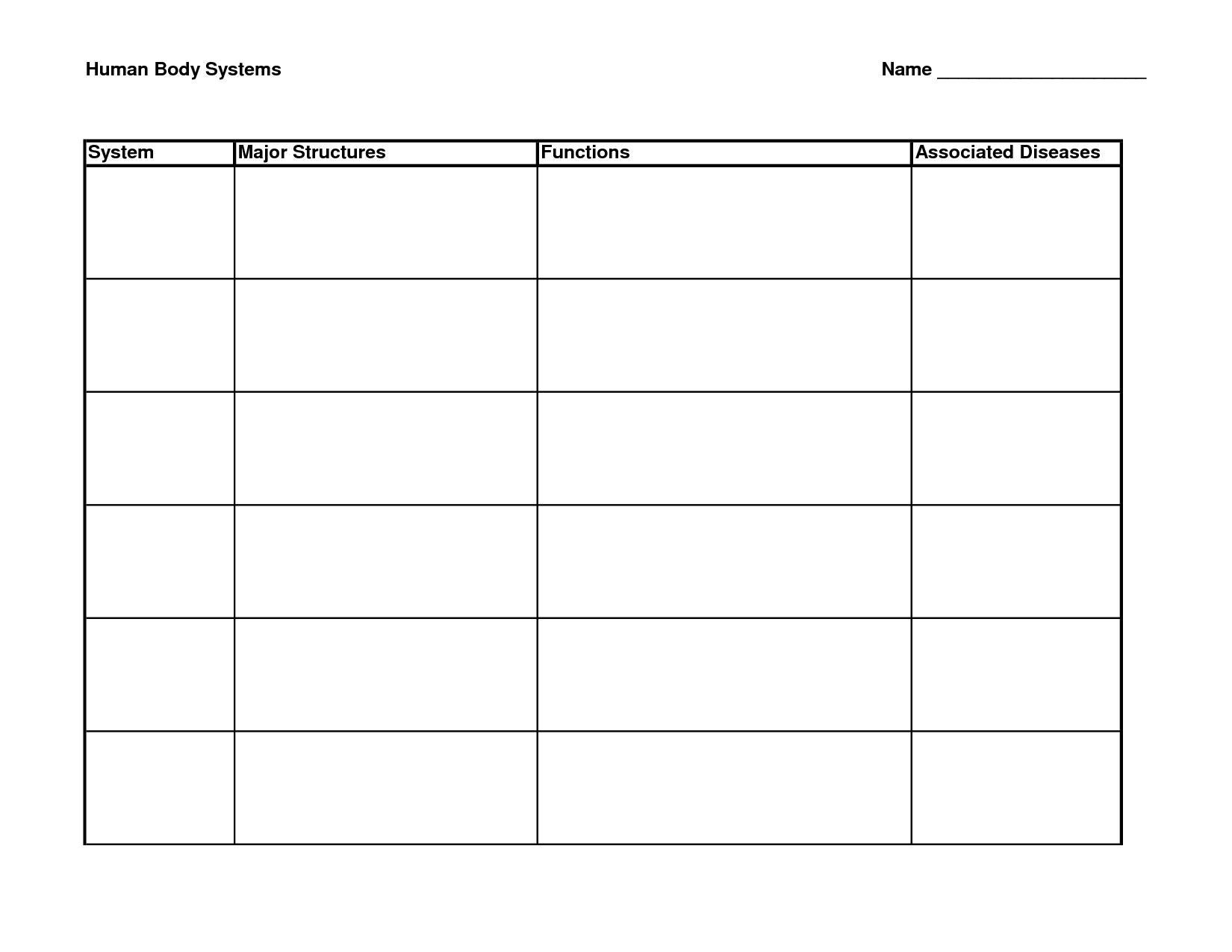
- Body System Worksheet Answer Key
- Anatomical Directional Terms Worksheet
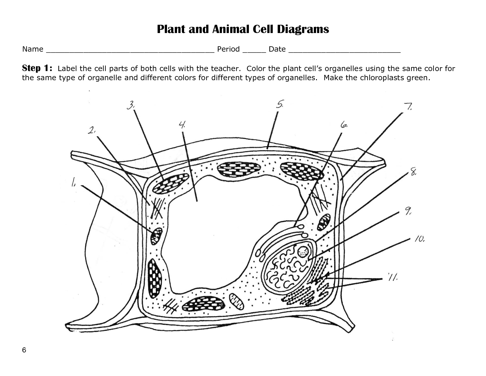
- Plant and Animal Cell Diagrams to Label
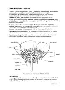
- Buttercup Flower Structure
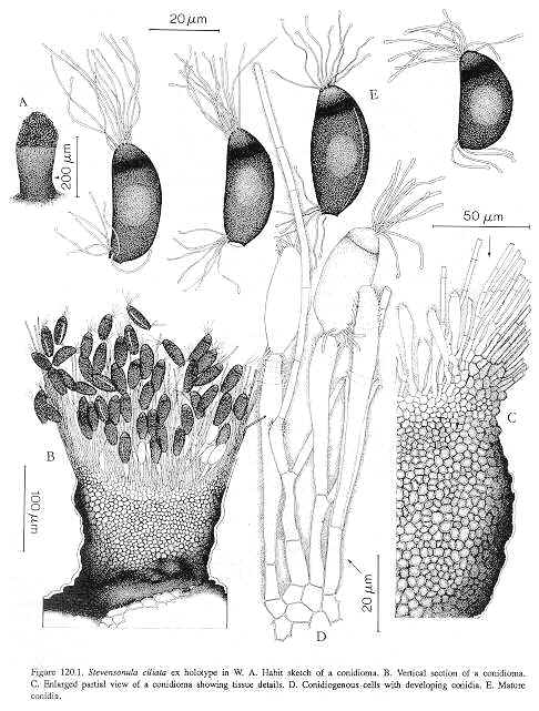
- Biological Drawings Line
- Microscope Slide Worksheet Blank
- Anime Girl Pencil Drawing Outline
- Anime Girl Pencil Drawing Outline
More Other Worksheets
Kindergarten Worksheet My RoomSpanish Verb Worksheets
Cooking Vocabulary Worksheet
My Shadow Worksheet
Large Printable Blank Pyramid Worksheet
Relationship Circles Worksheet
DNA Code Worksheet
Meiosis Worksheet Answer Key
Art Handouts and Worksheets
7 Elements of Art Worksheets
How is the heart structured?
The heart is a muscular organ that is divided into four chambers: the left atrium, left ventricle, right atrium, and right ventricle. These chambers are separated by valves that ensure blood flows in the correct direction. The heart is also surrounded by a double-layered sac called the pericardium, which helps protect and support the heart. Additionally, the heart has a network of blood vessels, including arteries and veins, that supply it with oxygen and nutrients while helping to distribute blood throughout the body.
What are the four chambers of the heart?
The four chambers of the heart are the left atrium, left ventricle, right atrium, and right ventricle.
What is the purpose of the atria?
The purpose of the atria is to receive blood from the veins and pass it on to the ventricles in the heart. They act as reservoirs that collect blood returning from the body or lungs and help propel it into the ventricles for efficient circulation throughout the body.
What is the purpose of the ventricles?
The ventricles in the brain function to produce and circulate cerebrospinal fluid, which acts as a cushion for the brain, provides nutrients, and removes wastes from the central nervous system.
What are the heart valves and what is their function?
The heart valves are the structures inside the heart that control the flow of blood by opening and closing at specific times during the cardiac cycle. There are four main heart valves - the tricuspid valve, pulmonary valve, mitral valve, and aortic valve. Their function is to ensure that blood flows in one direction through the heart chambers, preventing backflow and ensuring efficient circulation of oxygenated blood throughout the body.
What are the major blood vessels connected to the heart?
The major blood vessels connected to the heart are the aorta, pulmonary arteries, pulmonary veins, and vena cavae. The aorta carries oxygenated blood from the heart to the rest of the body, while the pulmonary arteries carry deoxygenated blood from the heart to the lungs for oxygenation. The pulmonary veins then return oxygenated blood from the lungs to the heart, and the vena cavae deliver deoxygenated blood from the body back to the heart.
What is the purpose of the coronary arteries?
The purpose of the coronary arteries is to supply oxygen-rich blood to the heart muscle. These blood vessels ensure that the heart receives essential nutrients and oxygen to function properly and maintain overall heart health.
How is blood flow regulated within the heart?
Blood flow within the heart is regulated by a combination of mechanisms, including the contraction and relaxation of the heart muscle (myocardium), the opening and closing of heart valves, and the coordination of electrical impulses that control the heart's activity. The cardiac conduction system, which includes the sinoatrial (SA) node, atrioventricular (AV) node, bundle of His, and Purkinje fibers, plays a crucial role in coordinating the rhythmic contractions of the heart chambers to ensure efficient blood flow throughout the body. Additionally, the adjustment of blood vessel diameter in response to changing demands, such as during exercise or stress, helps regulate blood flow within the heart and maintain circulation.
What is the role of the heart's electrical system?
The heart's electrical system is responsible for initiating and coordinating the heart's contractions. It generates electrical impulses that control the rhythm and rate of the heartbeat, ensuring that the heart pumps blood effectively throughout the body. This system includes structures like the SA node, AV node, and bundle of His, which work together to regulate the heart's activity and maintain a steady heartbeat.
How does the structure of the heart contribute to its overall function as a pump?
The structure of the heart, consisting of four chambers (two atria and two ventricles) separated by valves, plays a crucial role in its function as a pump. As blood enters the heart, it first fills the atria which contract to push the blood into the ventricles. The ventricles then contract to pump the blood out of the heart to the lungs and body. The valves ensure that blood flows in one direction, preventing backflow, while the muscular walls of the heart provide the necessary force to propel the blood through the circulatory system. This efficient structure allows the heart to continuously pump oxygen-rich blood to the organs and tissues of the body, supporting overall health and function.
Have something to share?
Who is Worksheeto?
At Worksheeto, we are committed to delivering an extensive and varied portfolio of superior quality worksheets, designed to address the educational demands of students, educators, and parents.











Comments