Simple Microscope Labeling Worksheet
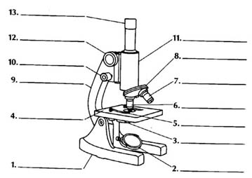
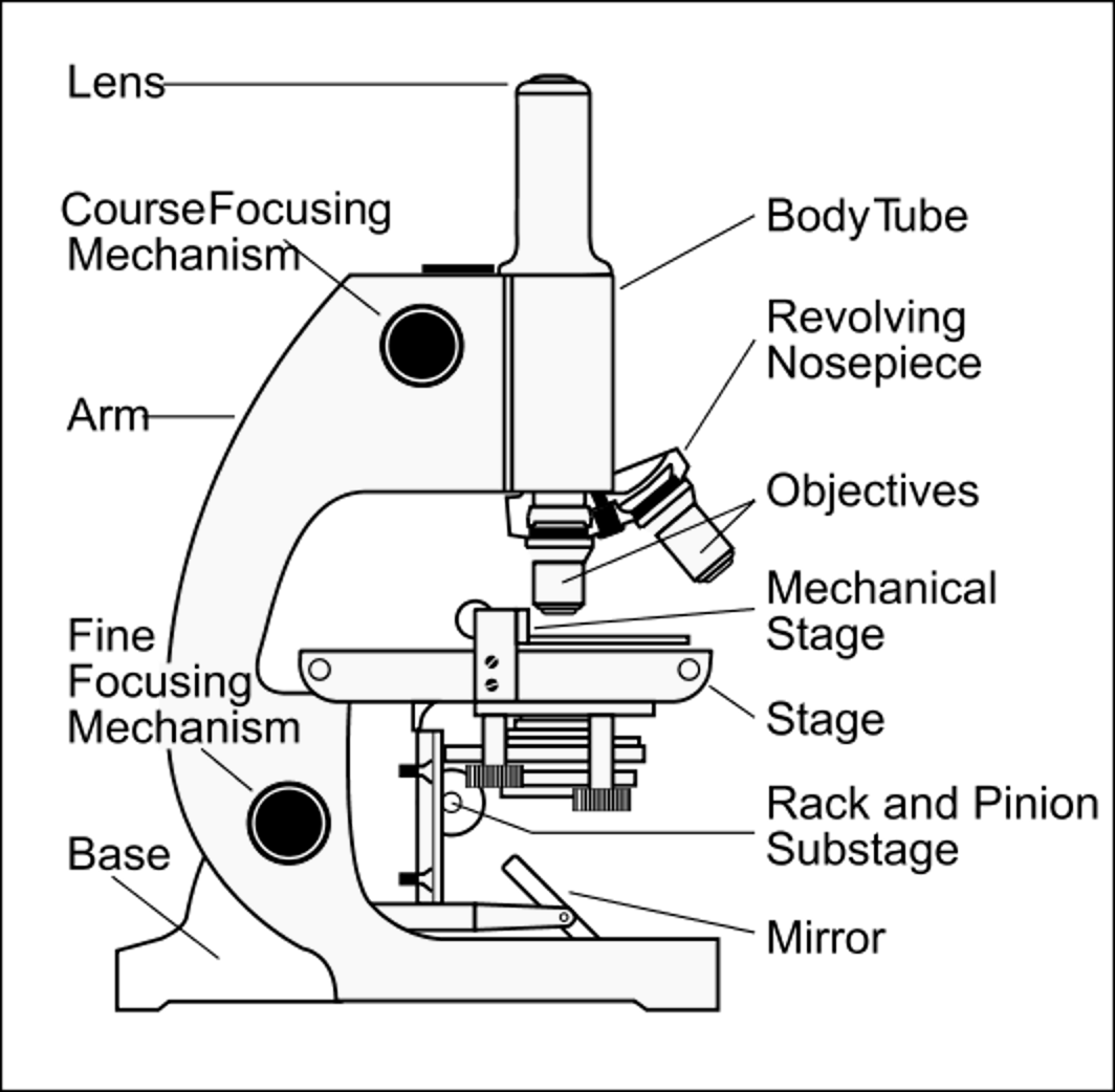
A simple microscope labeling worksheet is a useful tool for students who are learning about microscopes and the various parts that make up this essential scientific equipment. This worksheet aims to help students identify and understand the different components of a microscope, such as the eyepiece, objective lens, stage, and diaphragm. By engaging in this interactive activity, students can enhance their knowledge and comprehension of the microscope's entity and subject matter.
Table of Images 👆
- Compound Light Microscope Parts Blank
- Root Diagram Labeled
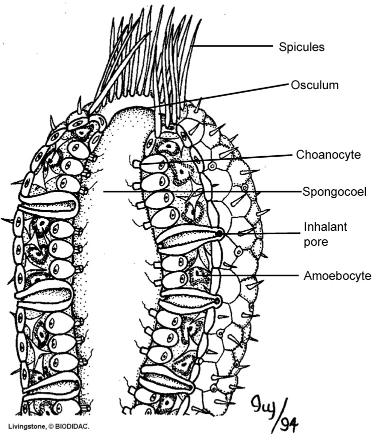
- Sponge Anatomy Diagram Answer Key
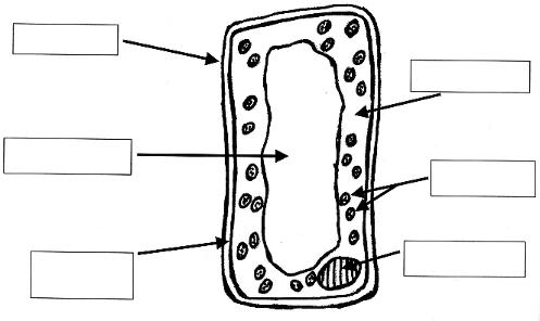
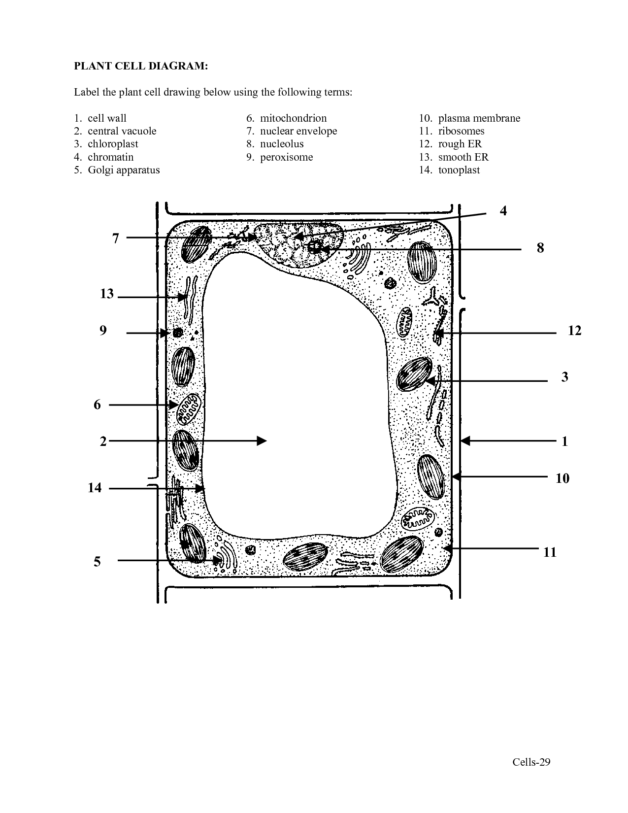
- Plant Cell Diagram without Labels
- Microscope Parts Labeled
- Spinal Cord Cross Section Diagram Labeled
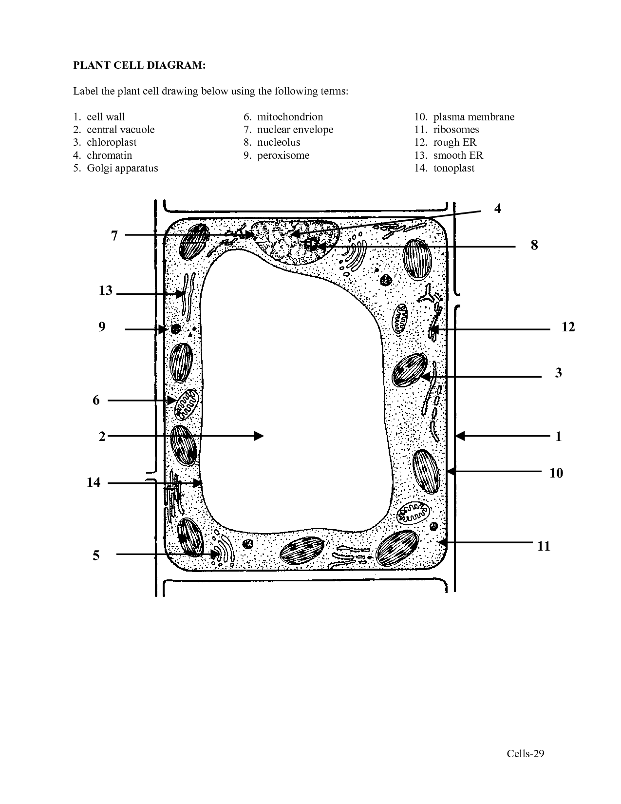
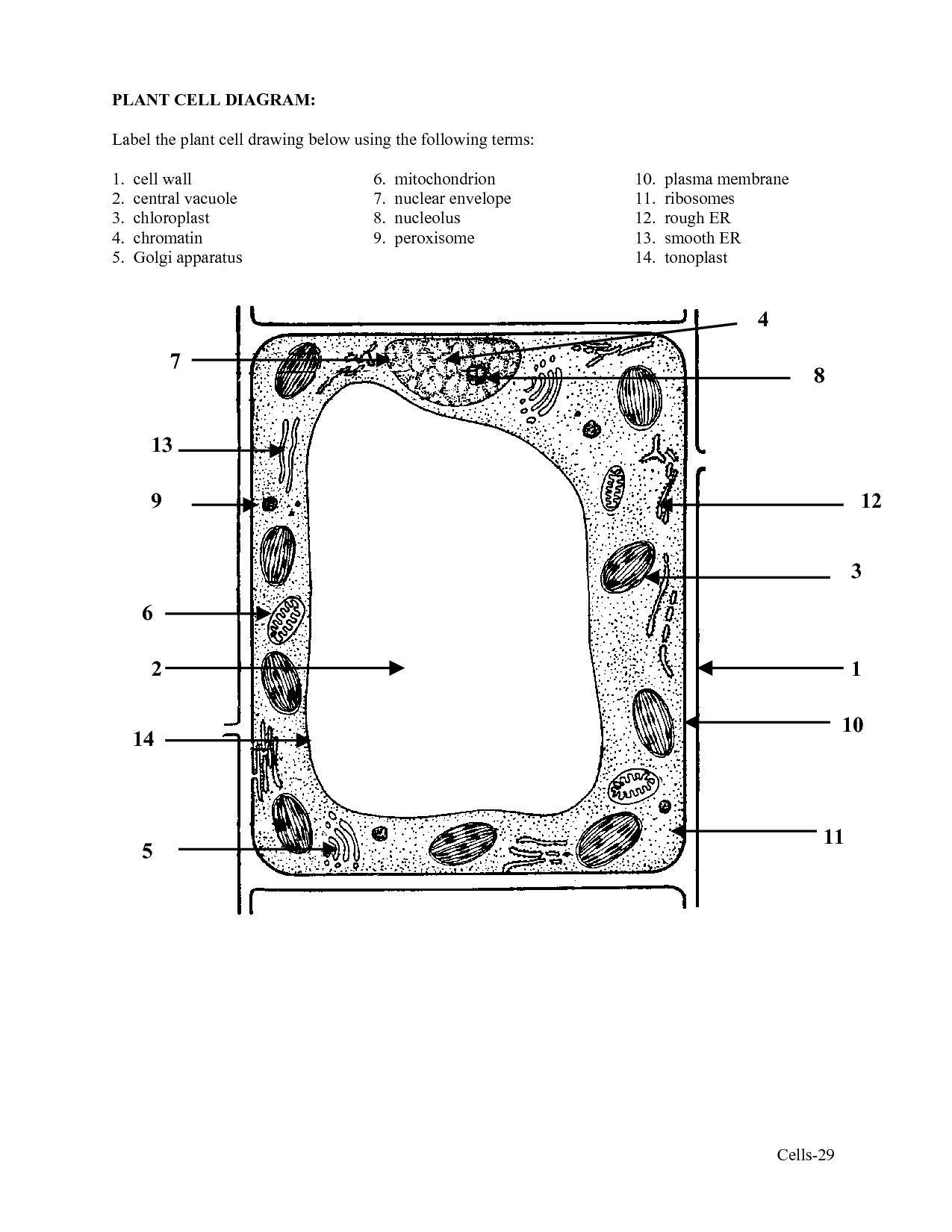
- Plant Cell Diagram with Labels
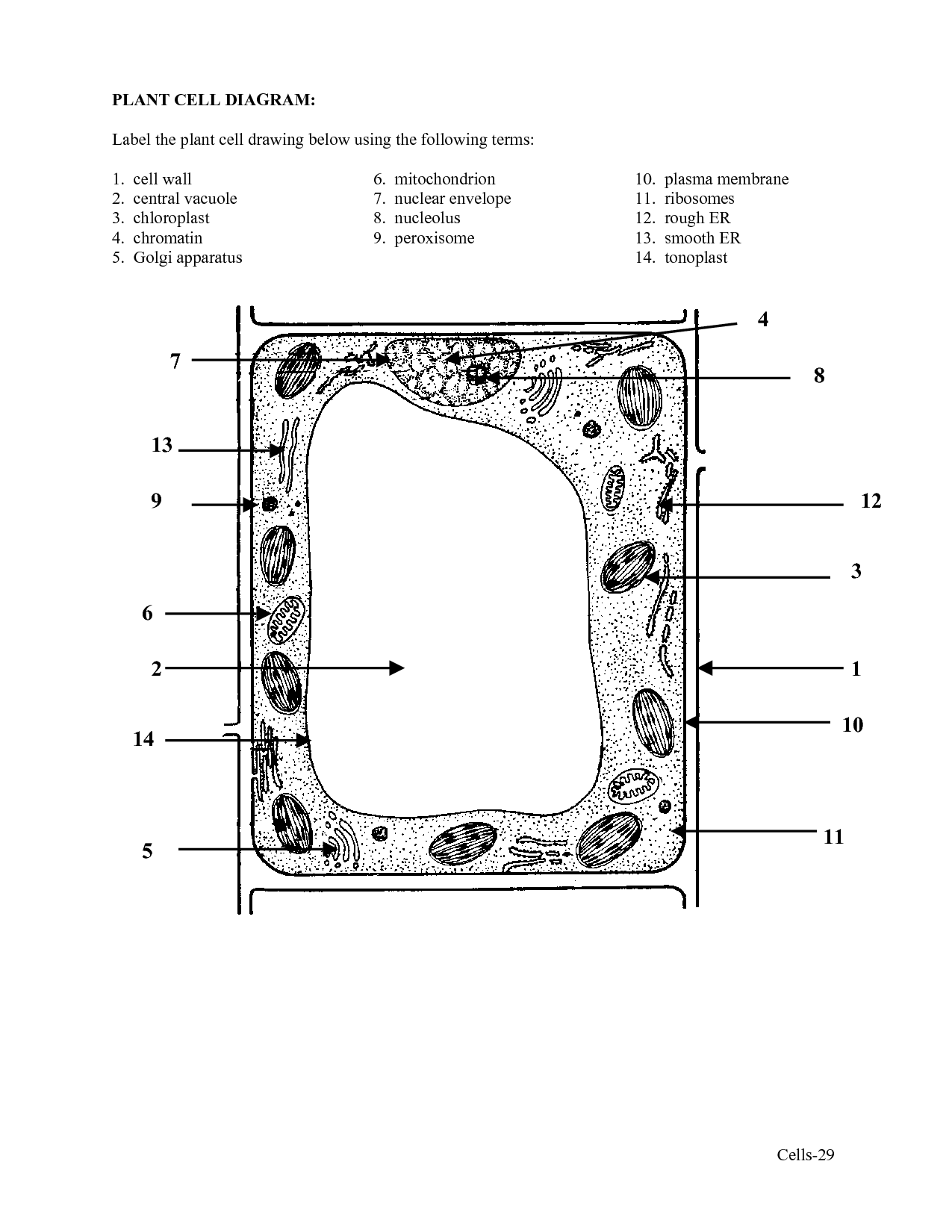
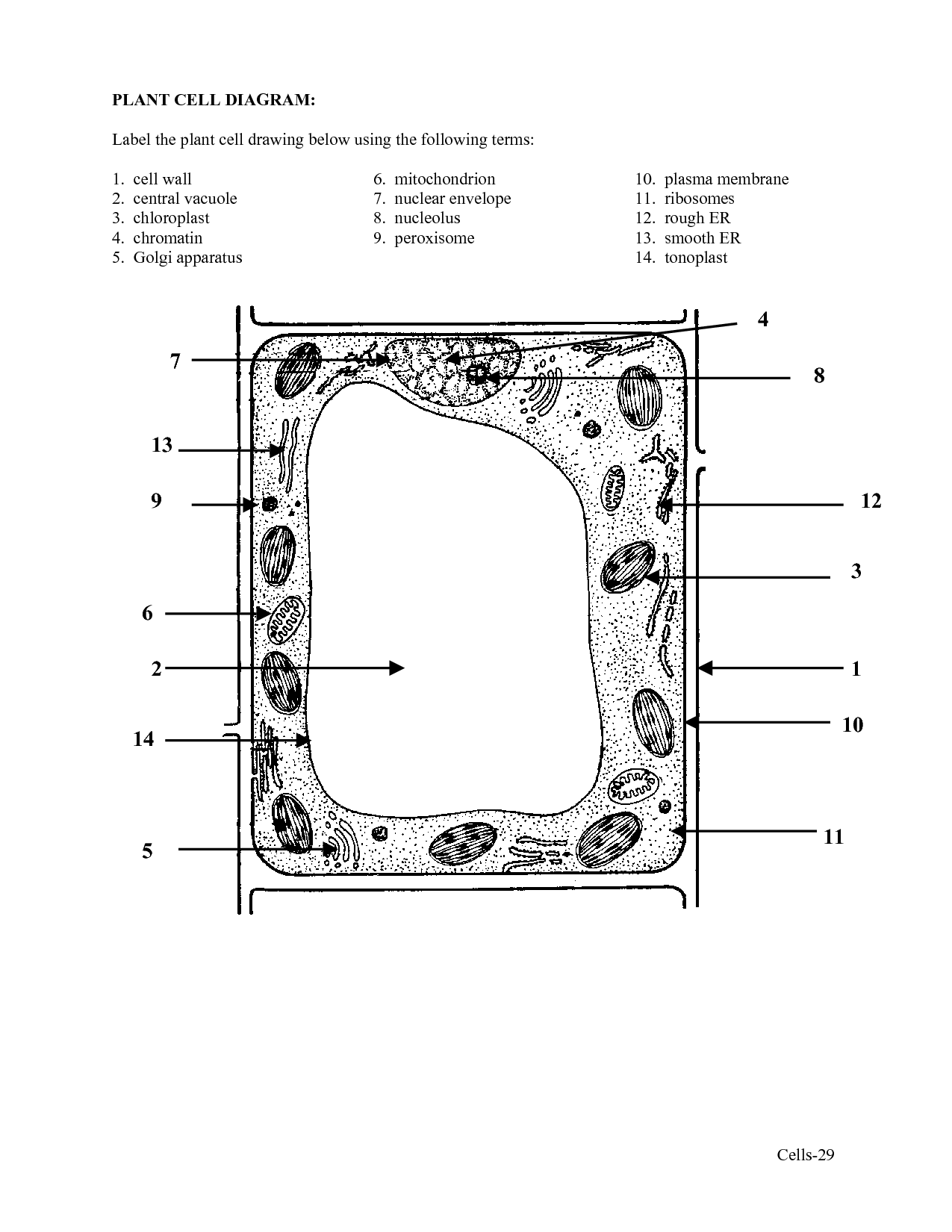
- Plant Cell Diagram with Labels
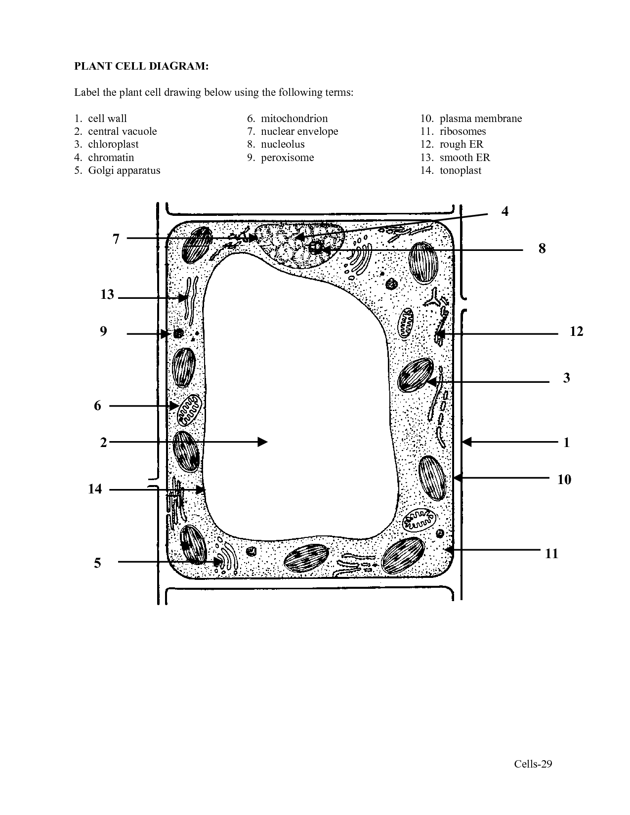
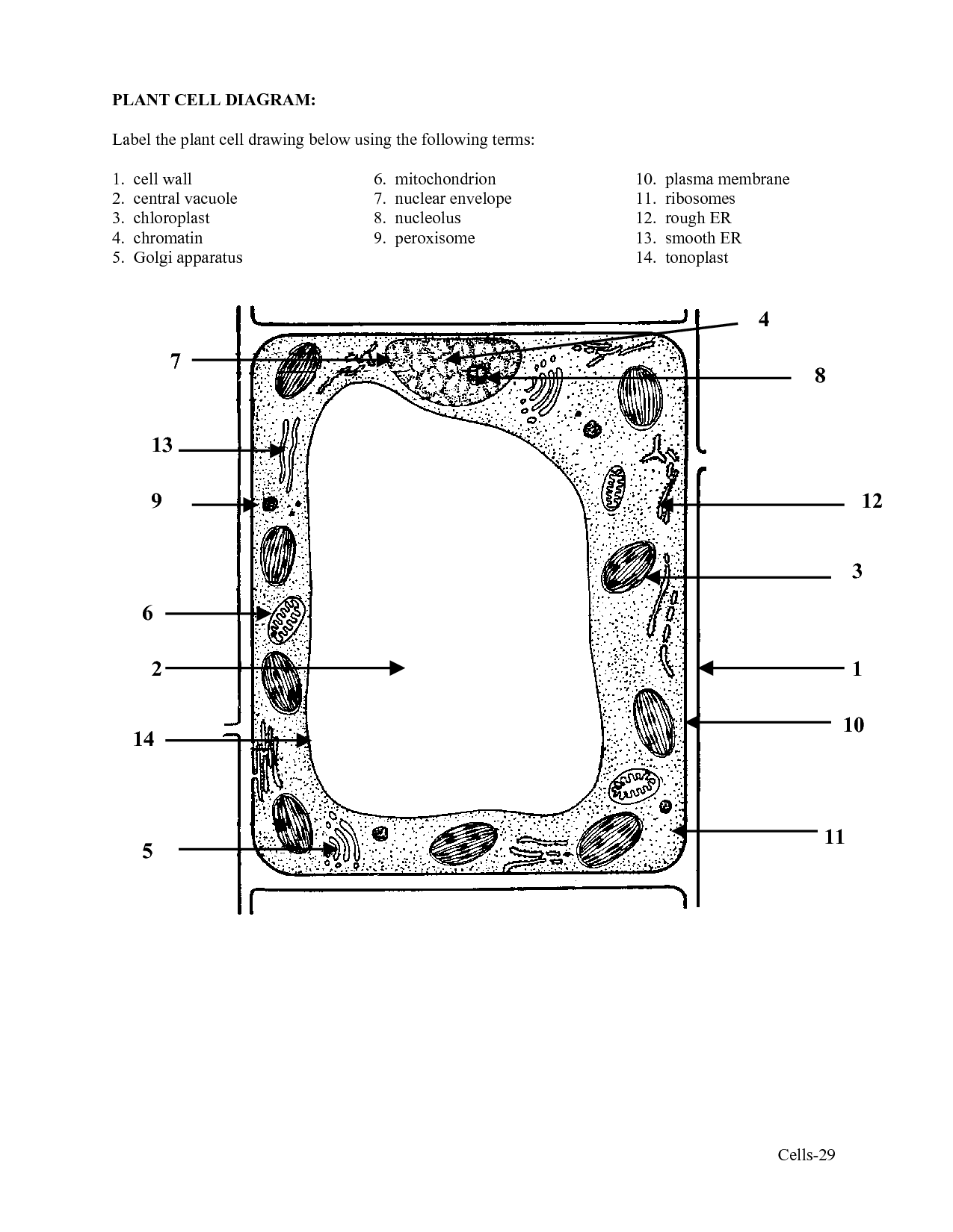
- Plant Cell Diagram with Labels
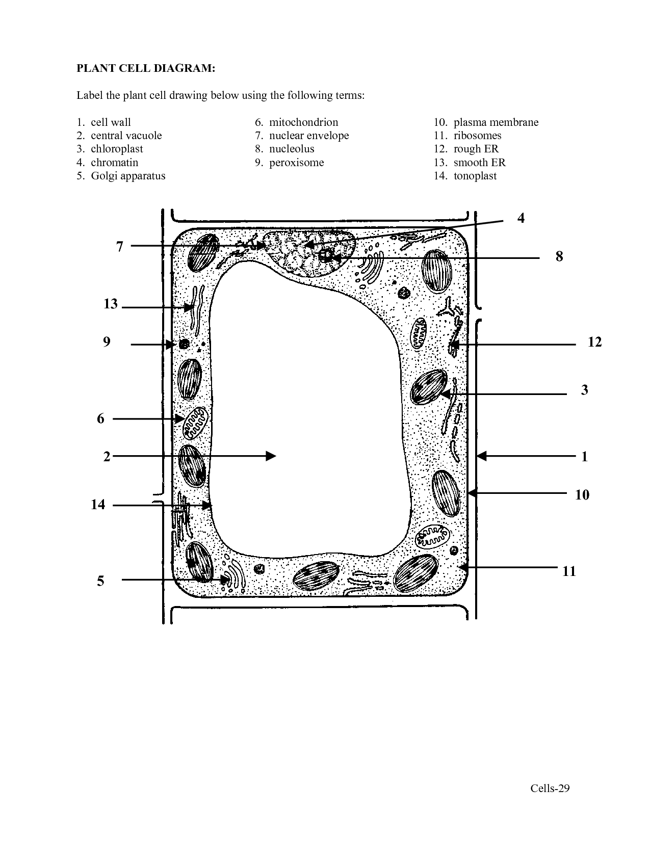
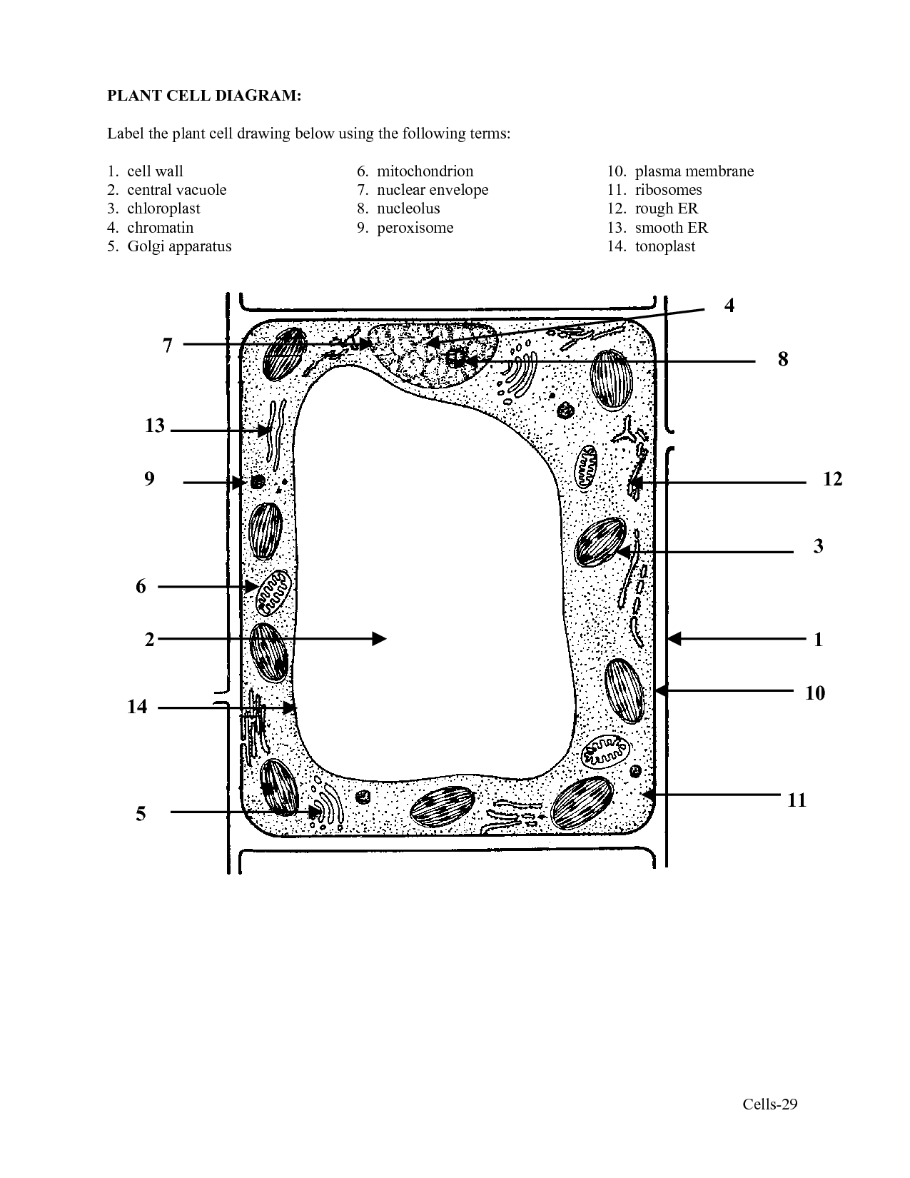
- Plant Cell Diagram with Labels
- Plant Cell Diagram with Labels
- Plant Cell Diagram with Labels
- Plant Cell Diagram with Labels
- Plant Cell Diagram with Labels
- Plant Cell Diagram with Labels
- Plant Cell Diagram with Labels
- Plant Cell Diagram with Labels
More Other Worksheets
Kindergarten Worksheet My RoomSpanish Verb Worksheets
Healthy Eating Plate Printable Worksheet
Cooking Vocabulary Worksheet
My Shadow Worksheet
Large Printable Blank Pyramid Worksheet
Relationship Circles Worksheet
DNA Code Worksheet
Meiosis Worksheet Answer Key
Rosa Parks Worksheet Grade 1
What is the purpose of a simple microscope?
The purpose of a simple microscope is to magnify small objects or specimens for closer observation and examination. It is a basic optical instrument consisting of a single lens that bends light rays to create a magnified image. Simple microscopes are often used in educational settings, laboratories, and scientific research to study the details of objects that are too small to be seen with the naked eye.
What are the main parts of a simple microscope?
A simple microscope typically consists of a single converging lens or a combination of lenses to magnify an object, an adjustable stage to hold the specimen, a light source to illuminate the specimen, and an eyepiece for the observer to view the magnified image. These basic parts work together to allow the user to see small objects more clearly by magnifying them.
What is the function of the eyepiece?
The function of the eyepiece in a microscope is to magnify the image produced by the objective lens, allowing the viewer to see a larger and more detailed image of the specimen being observed.
How does the objective lens magnify the image?
The objective lens magnifies the image by bending and focusing light rays from the object being observed. This bending of light rays allows the lens to bring the image into focus, making it appear larger and enabling the viewer to see fine details that would not be visible to the naked eye.
What is the role of the stage and stage clips?
The stage is where the specimen is placed for observation under the microscope, while stage clips are used to secure the slide in place on the stage to prevent it from moving during observation. The stage allows for precise positioning and movement of the specimen, enabling the viewer to focus on specific areas of interest, while the stage clips hold the slide securely to ensure that the specimen remains in place without shifting, ensuring accurate observations and data collection.
What is the purpose of the coarse adjustment knob?
The purpose of the coarse adjustment knob is to quickly move the stage up and down a significant distance to bring the specimen into focus initially.
What does the fine adjustment knob do?
The fine adjustment knob on a microscope is used to make precise and small changes to the focus of the specimen being observed. By turning the fine adjustment knob, you can bring specific details into sharp focus without making drastic changes to the overall focus of the image.
Where is the light source located in a simple microscope?
The light source in a simple microscope is typically located either above or below the specimen, usually as a built-in illumination system within the microscope itself.
How does the diaphragm control the amount of light?
The diaphragm in a camera controls the amount of light that reaches the camera sensor by adjusting the size of the aperture opening. By changing the size of the aperture, the diaphragm can either allow more or less light to pass through to the sensor, ultimately affecting the exposure of the photograph. A smaller aperture (higher f-stop number) lets in less light, resulting in a darker image, while a larger aperture (lower f-stop number) allows more light in, creating a brighter image.
What are the steps for properly using a simple microscope?
To properly use a simple microscope, first place the object you want to magnify on a clean and flat surface. Next, adjust the height of the lens so the object is in focus. Look through the eyepiece and use the focus wheel to sharpen the image further if needed. Move the object or the microscope to change the field of view or magnification. Finally, handle the microscope carefully and clean the lens with a soft cloth after use to maintain its quality.
Have something to share?
Who is Worksheeto?
At Worksheeto, we are committed to delivering an extensive and varied portfolio of superior quality worksheets, designed to address the educational demands of students, educators, and parents.







Comments