Human Eye Anatomy Diagram Worksheet
The human eye is a complex and fascinating organ that allows us to see the world around us. Understanding its anatomy is vital for anyone interested in biology, medicine, or optometry. This eye anatomy diagram worksheet provides a clear and concise overview of the different parts and structures of the human eye, making it an ideal resource for students and enthusiasts seeking to enhance their knowledge on this subject.
Table of Images 👆
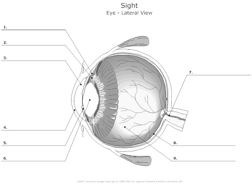
- Human Eye Diagram Labeled
- Cow Eye Dissection Worksheet Answer Key
- Parts of the Human Eye Diagram
- Human Eye Diagram Labeled
- Human Eye Diagram Unlabeled
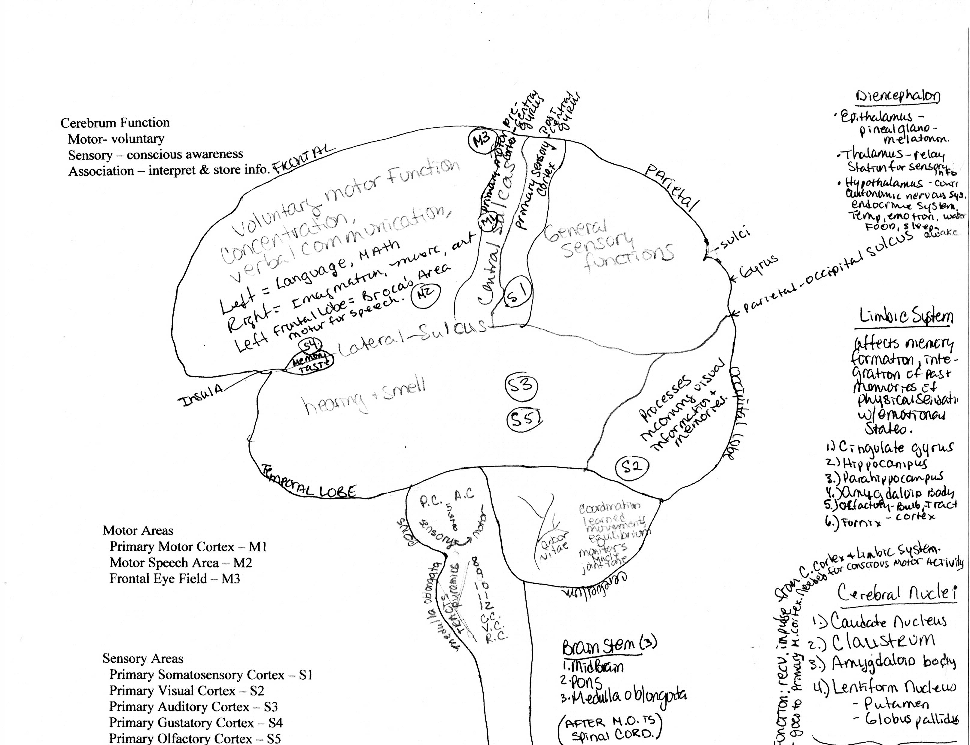
- Brain Anatomy Worksheet
- Kidney Nephron Diagram Unlabeled
- Polar Bear Skin Diagram
- Adjectives Describing Giant Pandas
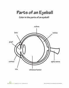
- Parts of the Eye Worksheet for Kids
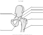
- Hip-Joint Anatomy Unlabeled
More Other Worksheets
Kindergarten Worksheet My RoomSpanish Verb Worksheets
Healthy Eating Plate Printable Worksheet
Cooking Vocabulary Worksheet
My Shadow Worksheet
Large Printable Blank Pyramid Worksheet
Relationship Circles Worksheet
DNA Code Worksheet
Meiosis Worksheet Answer Key
Rosa Parks Worksheet Grade 1
What is the function of the cornea?
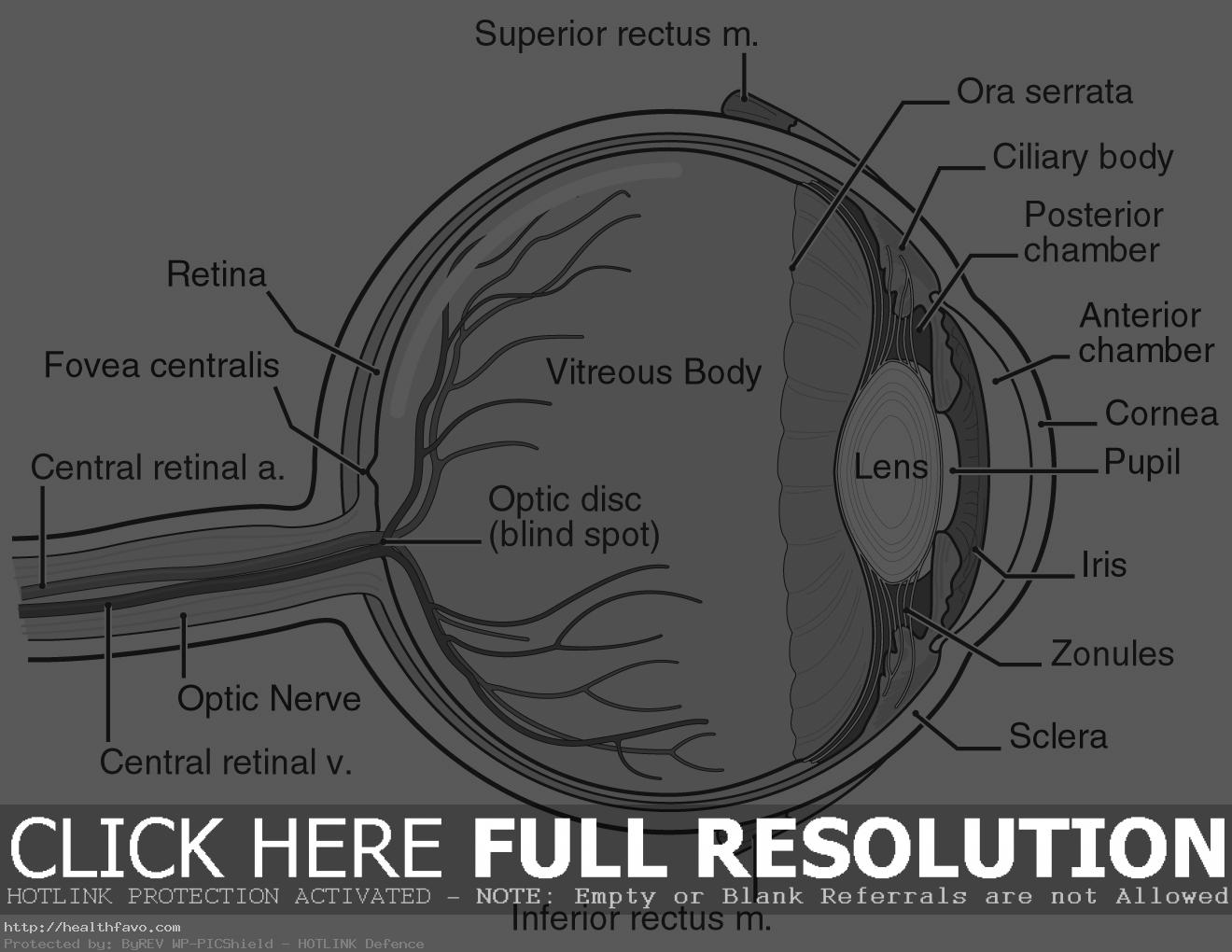
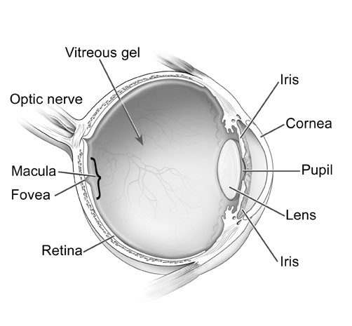
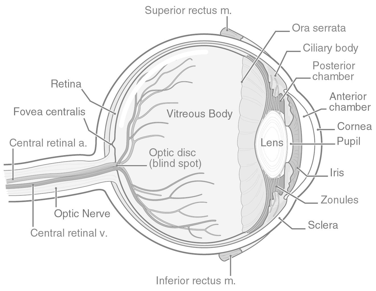
The cornea is the clear, dome-shaped outer surface of the eye that helps to focus light as it enters the eye. It plays a significant role in controlling and bending light rays to help produce a clear image on the retina, which is essential for clear vision.
What is the purpose of the iris in the eye?
The purpose of the iris in the eye is to control the amount of light entering the eye by adjusting the size of the pupil. This helps in regulating the amount of light that reaches the retina, which improves vision in different lighting conditions.
How does the lens contribute to vision?
The lens of the eye plays a crucial role in vision by focusing light onto the retina. It does this by changing its shape to adjust the focal point, allowing us to see objects at various distances clearly. This process, known as accommodation, is essential for the eye to create sharp images of the world around us.
What is the role of the retina in the eye?
The retina is a layer of tissue located at the back of the eye that consists of light-sensitive cells called photoreceptors. These photoreceptors detect light and convert it into electrical signals that are sent to the brain through the optic nerve. This process allows the brain to interpret the signals as visual images, thereby enabling us to see and perceive the world around us. Additionally, the retina also plays a crucial role in color perception, depth perception, and the ability to see in various lighting conditions.
How does the optic nerve assist in visual perception?
The optic nerve plays a critical role in visual perception by transmitting visual information from the eye to the brain. It carries electrical impulses generated by the photoreceptor cells in the retina to the visual processing areas in the brain, where the information is interpreted and processed to form a coherent visual perception. In essence, the optic nerve serves as the vital link between the eyes and the brain, allowing us to see and make sense of the world around us.
What is the function of the ciliary muscles?
The ciliary muscles, located in the eyes, play a crucial role in adjusting the shape of the lens to control the focusing ability of the eye. By contracting and relaxing, the ciliary muscles help to change the thickness of the lens, allowing the eye to focus on objects at different distances, a process known as accommodation.
What is the purpose of the aqueous humor in the eye?
The aqueous humor in the eye serves to provide nourishment to the avascular structures of the eye, such as the cornea and the lens, and helps maintain the intraocular pressure necessary for the eye to maintain its shape and function properly. Additionally, the aqueous humor also helps remove metabolic waste and protects the eye from harmful substances by assisting in the immune response of the eye.
How does the vitreous humor support eye structure?
The vitreous humor supports the structure of the eye by maintaining the shape of the eyeball, providing rigidity that helps hold the retina and lens in place, and transmitting light to the retina for vision. Additionally, it acts as a shock absorber, protecting the fragile structures inside the eye from impact or sudden movements.
What role do the eyelids play in protecting the eyeball?
Eyelids primarily act as a physical barrier to protect the eyeball from foreign objects, dust, and debris. They also help distribute tears to keep the surface of the eye moisturized and clean, promoting overall eye health and preventing dryness. Additionally, eyelids play a crucial role in blinking, which helps in spreading tears over the surface of the eye and removing any irritants that may have entered.
How does the tear duct system aid in maintaining eye health?
The tear duct system aids in maintaining eye health by producing and distributing tears to keep the eyes moist and lubricated. Tears help to wash away debris, dust, and foreign substances that can cause irritation or infection. Additionally, tears contain enzymes and antibodies that protect the eyes from microbial infections, adding a layer of defense against harmful pathogens. The tear duct system also plays a vital role in maintaining clear vision by ensuring the eyes are properly hydrated and nourished.
Have something to share?
Who is Worksheeto?
At Worksheeto, we are committed to delivering an extensive and varied portfolio of superior quality worksheets, designed to address the educational demands of students, educators, and parents.







Comments