E Microscope Worksheet Answers
Worksheets are a valuable learning tool that can enhance understanding and retention of complex subjects. Whether you're a student looking to reinforce your knowledge or a teacher seeking to engage your students, worksheets provide a structured and interactive approach to learning.
Table of Images 👆
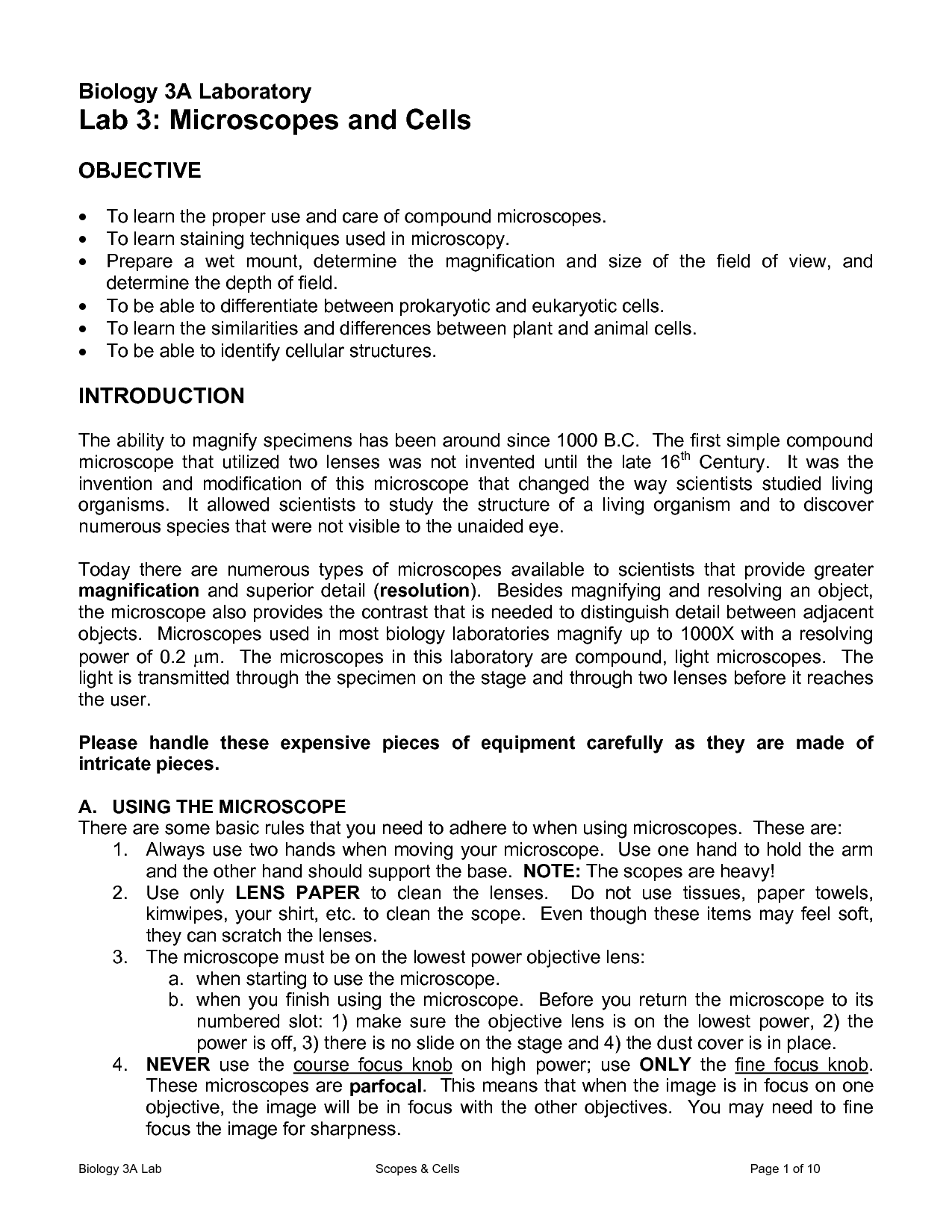
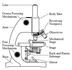
- Microscope Lab Worksheet Answers
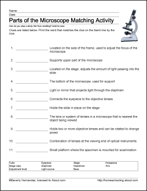
- Microscope Parts Worksheet Answers
- Microscope Parts Worksheet Crossword Puzzle Answers
- Light Microscope Parts Worksheet
- Printable Microscope Worksheet
- Microscope Lab Sheet for Kids
- Identifying Parts of a Microscope Worksheet
- Microscope Parts Printable Worksheet
- Microscope Labeling Quiz
- Electron Configuration Worksheet
- Light Microscope Parts and Functions
- Virtual Cell Worksheet Answer Key
- Microscope for Biology Worksheet Answers
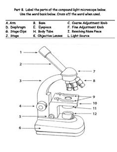
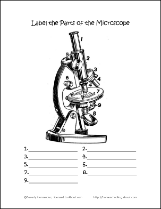
- Microscope Parts Blank Diagram
More Other Worksheets
Kindergarten Worksheet My RoomSpanish Verb Worksheets
Healthy Eating Plate Printable Worksheet
Cooking Vocabulary Worksheet
My Shadow Worksheet
Large Printable Blank Pyramid Worksheet
Relationship Circles Worksheet
DNA Code Worksheet
Meiosis Worksheet Answer Key
Rosa Parks Worksheet Grade 1
What is an electron microscope?
An electron microscope is a type of microscope that uses a beam of electrons to create a magnified image of a sample. It has a higher resolution and magnification capability compared to traditional light microscopes, allowing scientists to see tiny details of structures at the nanoscale level.
How does an electron microscope work?
An electron microscope works by using a beam of electrons instead of light to generate magnified images of samples. The electrons are accelerated through a vacuum towards the sample, and their interactions with the sample produce signals that are detected and used to create an image with much higher resolution than a traditional light microscope. The use of electrons allows electron microscopes to achieve much higher magnification and resolution than optical microscopes, making them essential tools in various scientific fields for studying the fine details of materials at the nanoscale level.
What are the advantages of using an electron microscope over a light microscope?
Electron microscopes offer higher magnification, resolution, and contrast compared to light microscopes. They can visualize smaller objects in greater detail due to the shorter wavelength of electrons, providing clearer images of tiny structures such as viruses, cellular organelles, and even individual atoms. Additionally, electron microscopes can capture images in 3D, revealing intricate details of biological and inorganic samples.
What types of samples can be studied using an electron microscope?
Electron microscopes are commonly used to study a wide range of samples including cells, tissues, microorganisms, nanoparticles, crystals, metals, ceramics, and polymers. These microscopes have the ability to provide high-resolution images and detailed information on the structure and composition of the samples, making them valuable tools in various fields such as biology, material science, nanotechnology, and geology.
How is the resolution of an electron microscope determined?
The resolution of an electron microscope is determined by the wavelength of the electrons being used. The shorter the wavelength, the higher the resolution. This is because shorter wavelengths can distinguish between smaller differences in structures, allowing for finer details to be observed in the specimen being studied. Other factors such as the quality of the lenses, the stability of the instrument, and the contrast mechanisms used also play a role in determining the overall resolution of an electron microscope.
What is the maximum magnification achievable with an electron microscope?
The maximum magnification achievable with an electron microscope is typically around 2 million times, although some advanced models can achieve magnifications up to 50 million times. This high level of magnification allows researchers to observe structures at the atomic level, making electron microscopes a powerful tool for studying materials and biological samples in great detail.
How does the use of electron beams in electron microscopes affect the sample being studied?
When electron beams are used in electron microscopes, they interact with the sample by penetrating the surface and creating signals such as secondary electrons, backscattered electrons, and X-rays. These interactions can alter the sample's surface by causing damage or charging effects. Additionally, the high-energy electron beams can generate heat and radiation, potentially affecting the sample's structure or composition. Therefore, careful consideration must be made in terms of beam energy, exposure time, and sample preparation to minimize any undesirable effects on the sample being studied.
What are the main components of an electron microscope?
The main components of an electron microscope include an electron source (such as a tungsten filament or field emission gun), electromagnetic lenses for focusing the electron beam, a specimen stage to hold and position the sample, detectors to capture the signals generated by interactions with the specimen, and a display or camera for viewing and recording the images produced by the microscope.
How are images generated in an electron microscope?
Images are generated in an electron microscope by focusing a beam of electrons onto the sample, which interacts with the atoms in the sample to produce signals such as backscattered electrons, secondary electrons, and characteristic X-rays. These signals are then detected and converted into an image by an electron detector, providing detailed information on the sample's structure and composition at a much higher resolution than traditional light microscopes.
What are some applications of electron microscopes in various fields of science and research?
Electron microscopes are widely used in various fields of science and research for their high resolution imaging capabilities. In biology, electron microscopes are used to study the detailed structure of cells and tissues, allowing scientists to better understand cellular functions and disease processes. In material science, electron microscopes are used to analyze the atomic structure of materials, helping researchers develop new materials with specific properties. In nanotechnology, electron microscopes are crucial for examining structures at the nanoscale level, paving the way for advancements in nanomaterials and nanodevices. Additionally, electron microscopes are used in fields such as geology, forensic science, and environmental science for a wide range of applications.
Have something to share?
Who is Worksheeto?
At Worksheeto, we are committed to delivering an extensive and varied portfolio of superior quality worksheets, designed to address the educational demands of students, educators, and parents.








Comments