Blank Microscope Worksheet
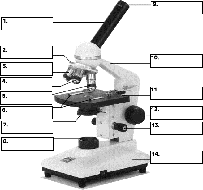
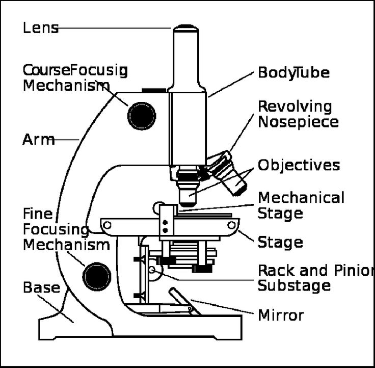
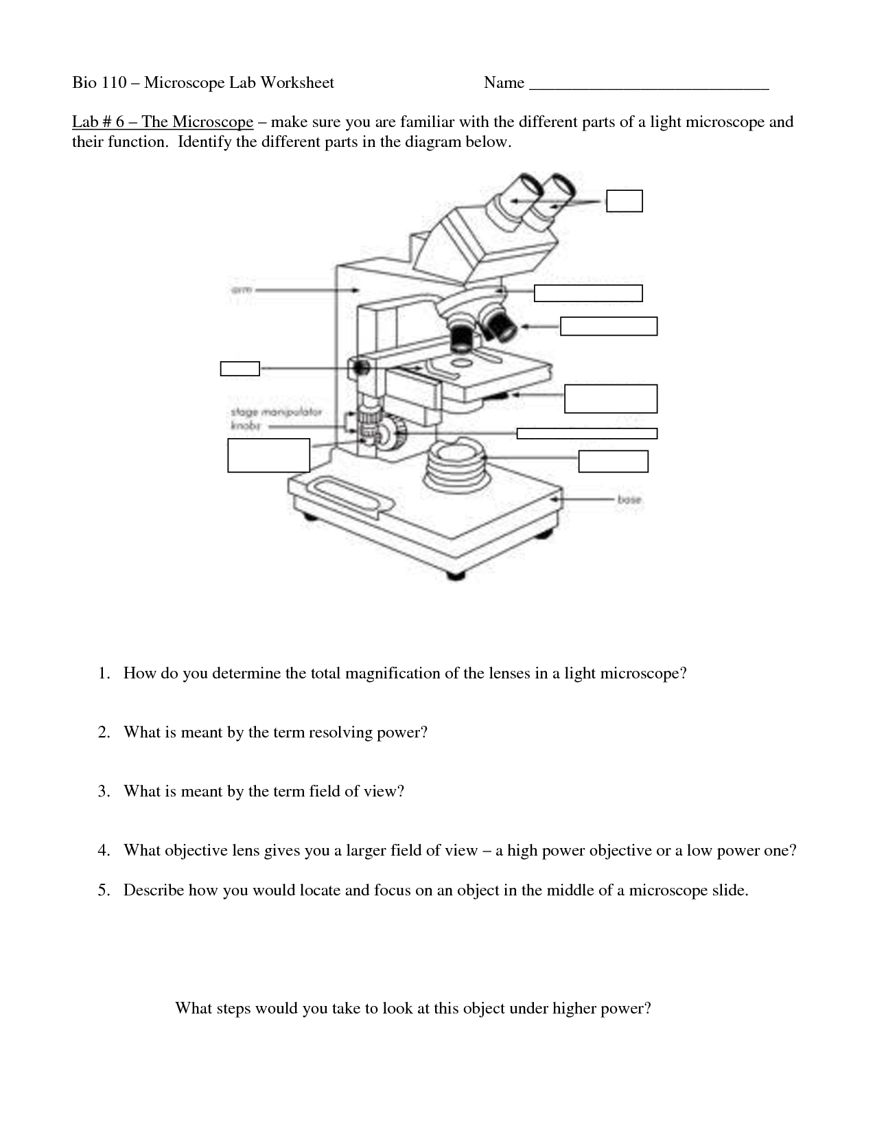
Are you currently seeking a comprehensive and user-friendly worksheet on microscopes for your science class? Look no further! We have the perfect solution for you. Our blank microscope worksheet is specifically designed to help students understand the various parts and functions of a microscope while engaging them in hands-on learning.
Table of Images 👆
- Blank Microscope Diagram
- Microscope Parts Quiz
- Human Heart Diagram Unlabeled
- Microscope Parts Worksheet
- Lord Hear Our Prayers Clip Art
- Compound Microscope Worksheet
- Blank Domino Addition Worksheet
- Labeled Flatworm Diagram
- Flat Stanley Template Printable
- Flat Stanley Template Printable
- Flat Stanley Template Printable
- Flat Stanley Template Printable
- Flat Stanley Template Printable
- Flat Stanley Template Printable
- Flat Stanley Template Printable
- Flat Stanley Template Printable
- Flat Stanley Template Printable
More Other Worksheets
Kindergarten Worksheet My RoomSpanish Verb Worksheets
Healthy Eating Plate Printable Worksheet
Cooking Vocabulary Worksheet
My Shadow Worksheet
Large Printable Blank Pyramid Worksheet
Relationship Circles Worksheet
DNA Code Worksheet
Meiosis Worksheet Answer Key
Rosa Parks Worksheet Grade 1
What is the purpose of a microscope?
The purpose of a microscope is to allow users to observe small objects and details that are not visible to the naked eye by magnifying them. This enables scientists, researchers, and students to study and analyze the structure, composition, and behavior of microscopic organisms and materials, leading to discoveries and advancements in various fields such as biology, medicine, geology, and material sciences.
What is the difference between a compound microscope and a stereo microscope?
The main difference between a compound microscope and a stereo microscope is the way they magnify objects. A compound microscope uses a series of lenses to magnify microscopic specimens in 2D, while a stereo microscope uses two separate optical paths to provide a three-dimensional view of larger specimens at lower magnifications. Stereo microscopes are commonly used for tasks that require depth perception and manipulation of larger objects, such as dissection, circuit board inspection, and quality control in manufacturing.
How does a microscope magnify an object?
A microscope magnifies an object by using a combination of lenses to bend and focus light rays onto the object being viewed. This process allows for the object to be enlarged and viewed in more detail than with the naked eye. By adjusting the focus and magnification levels of the lenses, a microscope can provide a highly detailed and magnified image of the specimen under observation.
What is the function of the eyepiece (ocular lens)?
The function of the eyepiece (ocular lens) in a microscope is to magnify the image created by the objective lens. By providing additional magnification, the eyepiece allows for better visualization of the specimen being observed under the microscope.
What is the significance of the objective lenses?
Objective lenses are a crucial component of optical instruments such as microscopes and telescopes, as they are responsible for gathering and focusing light to produce a magnified image. The significance of objective lenses lies in their ability to determine the magnification and resolution of the final image, making them instrumental in observing and analyzing small details, structures, or objects that would otherwise be difficult to see with the naked eye. Additionally, objective lenses also play a key role in the quality of the image produced, impacting factors such as clarity, contrast, and depth of field.
How does one calculate the total magnification of a microscope?
To calculate the total magnification of a microscope, multiply the magnification power of the objective lens by the magnification power of the eyepiece lens. Total magnification = magnification of objective lens x magnification of eyepiece lens.
Explain the concept of resolution in microscopy.
Resolution in microscopy refers to the ability of a microscope to distinguish two closely spaced objects as separate entities. It is the minimum distance at which two points can be distinguished as distinct and is influenced by factors such as the wavelength of light used, numerical aperture of the lens, and the quality of the optics. Higher resolution microscopes can produce clearer and sharper images with finer details, allowing for better visualization and analysis of specimens. Improving resolution in microscopy techniques is crucial for studying small biological structures and understanding complex biological processes at a cellular and subcellular level.
What is the role of the condenser in a microscope?
The role of the condenser in a microscope is to focus and concentrate the light onto the specimen being observed. By adjusting the condenser, the amount and angle of light entering the microscope can be controlled, allowing for clearer and more detailed visualization of the specimen. This helps to improve the contrast and resolution of the image seen through the microscope.
What is the difference between brightfield and darkfield microscopy?
Brightfield microscopy is a technique where the specimen appears darker than the surrounding bright field, providing contrast for visualization of stained samples, while darkfield microscopy is a technique where the specimen is illuminated with oblique light, causing light to scatter off the specimen and only objects with different refractive indices are visible against a dark background, making it useful for observing live, unstained samples with high contrast.
How does one properly handle and care for a microscope?
To properly handle and care for a microscope, ensure it is kept in a clean and dry environment, covered when not in use, and stored securely. Always use two hands when carrying the microscope and never lift it by the stage or the eyepiece. Cleaning the lenses with a lens paper and solution intended for optics is crucial, while avoiding touching the lenses with bare hands. Regular maintenance, such as checking for loose parts and adjusting the focus when needed, will help extend the lifespan of the microscope.
Have something to share?
Who is Worksheeto?
At Worksheeto, we are committed to delivering an extensive and varied portfolio of superior quality worksheets, designed to address the educational demands of students, educators, and parents.


Comments